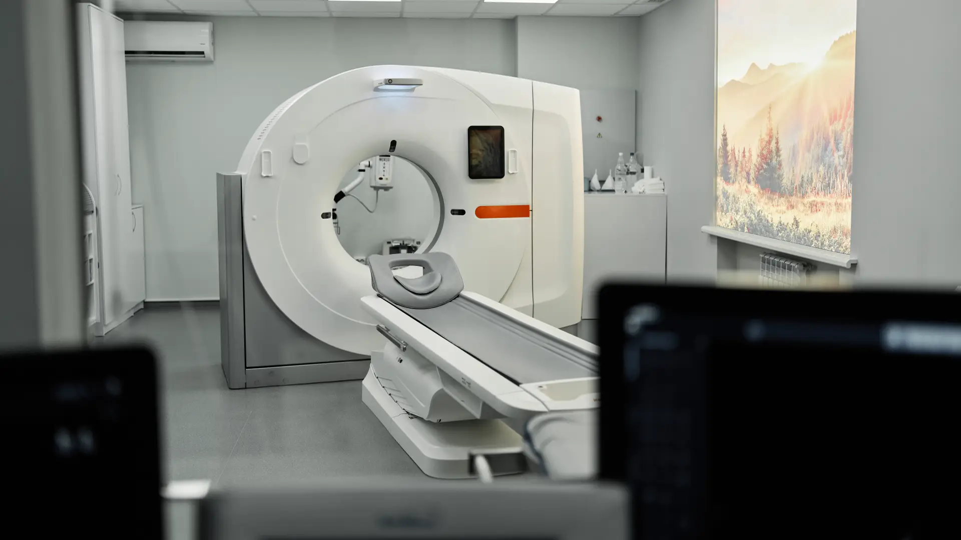
Radiology and Nuclear Medicine
Radiology and nuclear medicine cover all the imaging and treatment methods used in the diagnosis, staging and follow-up of cancer. Radiology shows the structure of organs; nuclear medicine provides information at the cellular and functional level. At ONCOMSI in Istanbul, these methods are assessed as part of an integrative treatment process coordinated by the medical oncologist.
İçindekiler
Toggle- What is nuclear medicine? A medical specialty that uses low doses of radioactive substances to show how organs work and, in some cases, to treat disease.
- Does PET/CT show cancer? It detects the increased metabolic activity of cancer cells and shows where the tumor is and whether it has spread.
- Which scan for which cancer? FDG PET/CT for lung cancer and lymphoma, PSMA PET/CT for prostate cancer, bone scintigraphy for suspected bone metastases.
- Who decides and interprets? At ONCOMSI, the medical oncologist chooses the test and interprets the result together with the patient’s full clinical picture.
What Are Radiology and Nuclear Medicine?
Nuclear medicine is the branch of medicine that uses low doses of radioactive substances (radiopharmaceuticals) to diagnose and treat disease. The substance collects in the organ being examined and is imaged with special cameras, so what is assessed is not the organ’s structure but how it works. Radiology, on the other hand, produces structural images of organs with methods such as MRI, CT, ultrasound and mammography. When the two fields are used together, the location of the cancer, its spread and the response to treatment can be assessed more precisely.
Radiologic Imaging Methods in Cancer
- Magnetic resonance imaging (MRI): shows soft tissue, the brain, the spine and abdominal organs in detail without radiation.
- Computed tomography (CT): assesses tumors, bleeding and vascular disease with cross-sectional images.
- Ultrasound and Doppler ultrasound: examine soft tissue and blood flow with sound waves.
- Mammography: used for the early detection of breast cancer.
- X-ray: allows quick assessment of bone and lung structures.
Nuclear Medicine Imaging: PET/CT, SPECT/CT and Scintigraphy
PET/CT combines the metabolic information of positron emission tomography with the anatomic map of computed tomography in a single device. Because cancer cells have a high metabolic rate, they take up more of the radiopharmaceutical; PET shows this activity and CT shows its location. Different radiopharmaceuticals are used: FDG for general oncologic imaging, Ga-68 PSMA for prostate cancer, Ga-68-labeled somatostatin analogs (such as DOTATATE) for neuroendocrine tumors, and choline and FAPI for specific situations.
SPECT/CT combines single-photon emission imaging with CT to locate findings in the bones, heart and thyroid. Scintigraphy is performed with a gamma camera: bone and thyroid scintigraphy is used to look for bone metastases and to assess thyroid disease. For the details of the PET/CT process, see how a PET/CT scan is done and how to prepare; for its types, see Ga-68 PSMA and other types of PET/CT.
Which Imaging Method for Which Cancer?
- Lung cancer and lymphoma: FDG PET/CT for spread and treatment response.
- Prostate cancer: Ga-68 PSMA PET/CT.
- Suspected bone metastasis: whole-body bone scintigraphy.
- Breast: mammography and, when needed, MRI.
- Thyroid: thyroid scintigraphy and radioactive iodine applications.
The choice of method is made by the medical oncologist, who interprets the imaging result together with the patient’s full clinical picture.
MRI, CT and Ultrasound: Which One and When?
- MRI: when soft-tissue detail is needed and when radiation should be avoided.
- CT: for fast, wide-area scanning and for assessing the lungs and bones.
- Ultrasound: as a radiation-free first step and for examining blood flow.
What Do the Imaging Result and the SUV Value Mean?
The SUV value in PET/CT reports shows how much of the radiopharmaceutical a tissue takes up, in other words its metabolic activity. High uptake alone does not mean a definite diagnosis; inflammatory conditions can also show uptake. For this reason, the result is interpreted by the medical oncologist together with the patient’s history, other tests and examination.
PET/CT is also used to follow the response to treatment. In a single case report published in 2026, FDG PET/CT showed a 63.7% metabolic response after one month of Metabolically Controlled Oncologic Therapy (MCOT) in a patient with drug-resistant advanced pancreatic cancer (İyikesici MS. Curr Oncol. 2026;33(8):489. PMID 42645394). As a single case, this finding cannot be generalized to other patients.
Nuclear Medicine Treatments and the Theranostic Approach
Radioactive iodine (I-131) therapy is used in thyroid disease and thyroid cancer; the substance, taken up by thyroid cells, acts in a targeted way. Theranostics combines “therapy” and “diagnostics”: the same target molecule is used both for imaging and for treatment. According to some approaches, prostate cancer imaged with Ga-68 PSMA may be treated with Lu-177 PSMA; this “see and treat” logic can also be applied in neuroendocrine tumors. For details, see theranostic treatment (Lu-177 PSMA).
Preparation Before the Test and Radiation Safety
In tests such as PET/CT, a waiting period is needed after the radiopharmaceutical is given, and then the imaging is performed. Preparation instructions (for example, fasting) vary with the type of test and are given by the physician in advance; in conditions such as diabetes, preparation is planned individually. The amount of radioactive substance is planned at low doses; pregnancy, breastfeeding and the situation of any accompanying person are assessed beforehand.
Radiology and Nuclear Medicine at ONCOMSI: The Patient Journey
Nuclear medicine and imaging in Istanbul is part of the integrative treatment process at ONCOMSI. The medical oncologist is the conductor of the whole process: deciding on the test, interpreting the result and managing the treatment. The patient journey follows the steps of initial consultation, planning, procedure and follow-up. Assoc. Prof. Mehmet Salih İyikesici and his specialist team stand by the patient at every step. Patients from every part of Istanbul and from abroad can access this process.
The Imaging Process for Patients Coming from Abroad
Istanbul is a center where international patients can easily access imaging and nuclear medicine tests. For patients coming from abroad, the process begins with a review of existing tests and reports before travel, so that unnecessary repeat tests can be avoided on arrival in Istanbul. At ONCOMSI, the imaging process for international patients is also coordinated by the medical oncologist, and the next steps are explained to the patient clearly. For more, see the international patients page.
Frequently Asked Questions
What is nuclear medicine and what is it used for?
Nuclear medicine is the branch of medicine that assesses both the structure and the function of organs using low doses of radioactive substances. It is used in the early diagnosis, staging and treatment follow-up of cancer, heart disease and thyroid disease.
Does PET/CT show cancer?
PET/CT shows the location and spread of a tumor by detecting the increased metabolic activity of cancer cells. It is a widely used imaging method for cancer diagnosis, staging and assessing the response to treatment.
What is the difference between PET/CT and SPECT/CT?
Both combine functional imaging with CT. PET/CT offers higher resolution with positron-emitting radiopharmaceuticals; SPECT/CT uses single-photon-emitting substances and is used in areas such as bone, heart and thyroid.
How is a PET/CT scan done?
A radiopharmaceutical is given through a vein, a waiting period is needed for it to distribute, and then imaging is performed in the scanner. The total time varies with the type of test and is communicated in advance.
What should I pay attention to before a PET/CT scan?
Preparation usually includes a fasting period and varies with the type of test. In conditions such as diabetes, preparation is planned individually. Exact instructions are given by your physician before the appointment.
Are nuclear medicine tests harmful?
The amount of radioactive substance used is planned at low doses, and procedures are carried out under specialist supervision. Pregnancy, breastfeeding and the situation of any accompanying person are assessed beforehand.
What is a Ga-68 PSMA PET/CT scan for?
Ga-68 PSMA PET/CT binds to PSMA (prostate-specific membrane antigen) on prostate cancer cells and shows the location and spread of the disease. Neuroendocrine tumors are imaged with different Ga-68-labeled molecules.
What is theranostic treatment?
Theranostics is the use of the same target molecule for both diagnosis and treatment. For example, according to some approaches, prostate cancer imaged with Ga-68 PSMA may be treated with Lu-177 PSMA. It is based on a “see and treat” logic.
What should I pay attention to after radioactive iodine therapy?
Radioactive iodine (I-131) therapy is used in thyroid disease and thyroid cancer. Isolation and daily-life recommendations after treatment are planned for each patient and explained by the specialist in advance.
Are nuclear medicine and medical oncology offered together in Istanbul?
At ONCOMSI in Istanbul, imaging and nuclear medicine are assessed as part of an integrative treatment process coordinated by the medical oncologist. Results are interpreted not in isolation but together with the patient’s full clinical picture.
Appointments and Contact
Contact us for an appointment. For your radiology and nuclear medicine tests and your treatment plan in Istanbul, you can receive an assessment at ONCOMSI coordinated by a medical oncologist. Reach us through the contact page.
This page is for informational purposes only. Please consult your doctor for diagnosis, treatment, and personal health decisions.